Biomedical engineers inspire research to rejuvenate stem cells for transplants

For more than 70 years, stem cell transplantation has proven an effective therapy for fighting blood and immune diseases. But the bone marrow blood cells most often used in transplants are not powerful as fetal blood stem cells. To boost the therapeutic abilities of adult stem cells, scientists seek the biological switches that could rejuvenate their ability to produce healthy blood cells and treat disease.
While adult blood stem cells reside in the bone marrow, new research from University of Illinois Chicago sought out these switches in an unusual location: the developing liver. There, they isolated a pathway that controls the potency of the blood-producing stem cells from mice, discovering that the key ingredient may be provided by the stem cells themselves.
The work, published in Proceedings of the National Academy of Sciences, advances a novel approach that looks to the environment where these hematopoietic stem cells naturally thrive for clues on how to unlock their medical applications.
“We’re seeking the location that provides us the best ability to learn about where stem cells naturally expand and grow,” said Kostandin Pajcini, senior author of the study and associate professor of pharmacology at UIC. “Surprisingly, as we studied this, we realized that it’s not the bone marrow, but the fetal liver.”
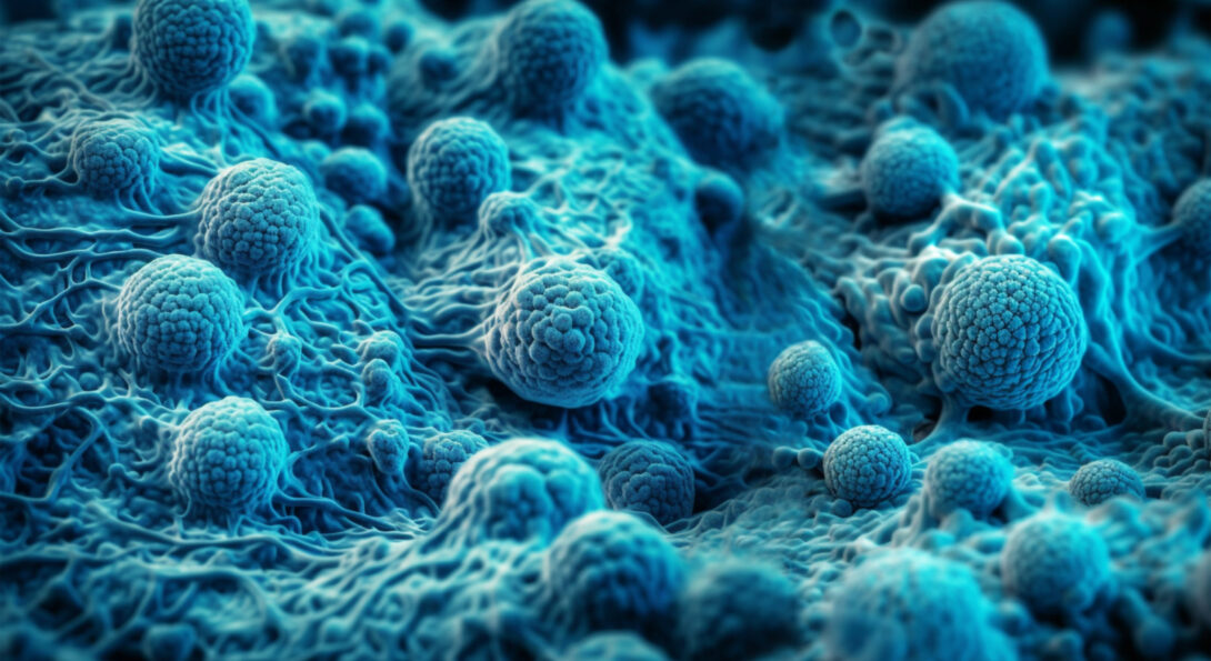
The therapeutic promise of stem cells lies in their unfettered possibilities. With the ability to become many different cell types, stem cells are critical for creating the organs, blood and structures of the developing body. In adulthood, the hematopoietic, or blood-producing, stem cells reside within the bone marrow and regularly activate to replenish the supply of red and white blood cells and platelets.
That unique ability also makes them a valuable tool for treating patients with cancer or blood disease. When the normal course of blood production is disrupted — for example, by chemotherapy or autoimmune dysfunction — a stem cell transplant can reboot the factory for the circulatory system.
But despite decades of successful stem cell transplants, the therapy remains unreliable and in high demand. Finding the right donor match can be difficult, and transplants sometimes fail to take hold. Scientists have tried to grow or rejuvenate stem cells in the laboratory to subsequently use in transplants but have struggled to find methods that boost their healing abilities.
While many laboratories have looked at the factors that activate hematopoietic stem cells in their adult home of the bone marrow, Pajcini’s group has focused on the fetal liver. During development, hematopoietic stem cells gather in the liver and burst into action, expanding the blood stem cells to circulate through a growing embryo and later populate the bone marrow.
“Stem cells have this unique lifestyle, where they’re like a bear hibernating in the bone marrow, and then they’re jumping around and dancing in the fetal liver,” Pajcini said.
One key difference between bone marrow and the fetal liver is the activity of the Notch pathway, a cellular signal well-studied for its importance in developmental biology. While Notch activity in the bone marrow stem cells is suppressed, keeping them largely dormant, it is elevated in fetal liver. But the trigger for that pathway, and the cell type that carries it, remained mysterious.
In the new paper, led by Dr. Lijian Shao, visiting instructor at UIC, researchers used single-cell analysis and genetic knockout mouse models to examine the role of jagged1, a protein that activates the Notch pathway. Blocking jagged1 and turning off the Notch pathway made stem cells from mice much less effective after transplants, while replacing the signal reignited their functionality.
But where, in the normal fetal liver, is this jagged1 activation coming from? Surprisingly, the researchers discovered that the “on” signal comes from the stem cells themselves.
“This was astounding and unexpected,” Pajcini said. “This result indicates that the stem cells are not so much reliant on the other cells in the fetal liver, but that they potentially rely on themselves and their progeny to activate the signaling in a cyclical fashion.”
This proximity effect could explain why stem cells are so much more lively in fetal liver than in bone marrow, where adult stem cells are largely isolated from each other. Adapting that phenomenon to create rejuvenated adult stem cells might require both cellular factors and an environment that keeps stem cells close together — a hypothesis Pajcini’s group is testing with 3D-printed models replicating the fetal liver tissue, created by Salman Khetani from the Richard and Loan Hill Department of Biomedical Engineering.
“In the fetal liver, stem cells don’t have a place to hide. They haven’t found a location where they’re going to go yet, so they’re much more active, more proliferative and also more functional,” Pajcini said. “Perhaps we can re-create a microenvironment that provides them with the right and proper signals to rejuvenate and reprogram them, using the process that’s already developmentally in place.”
In addition to Shao and Pajcini, paper co-authors included Na Yoon Paika, Mark A. Sanborn, Thilinie Bandara, Kilian Sottoriva and Dr. Jalees Rehman of UIC, and Anjali Vijaykumar and Cesar Nombela-Arrieta of University Hospital Zurich. The work was funded by NIH grants R01HL134971, R01HL151720, and UL1TR002003.