Shokuhfar lab publication in Small Methods journal
Small Methods Journal Article Heading link

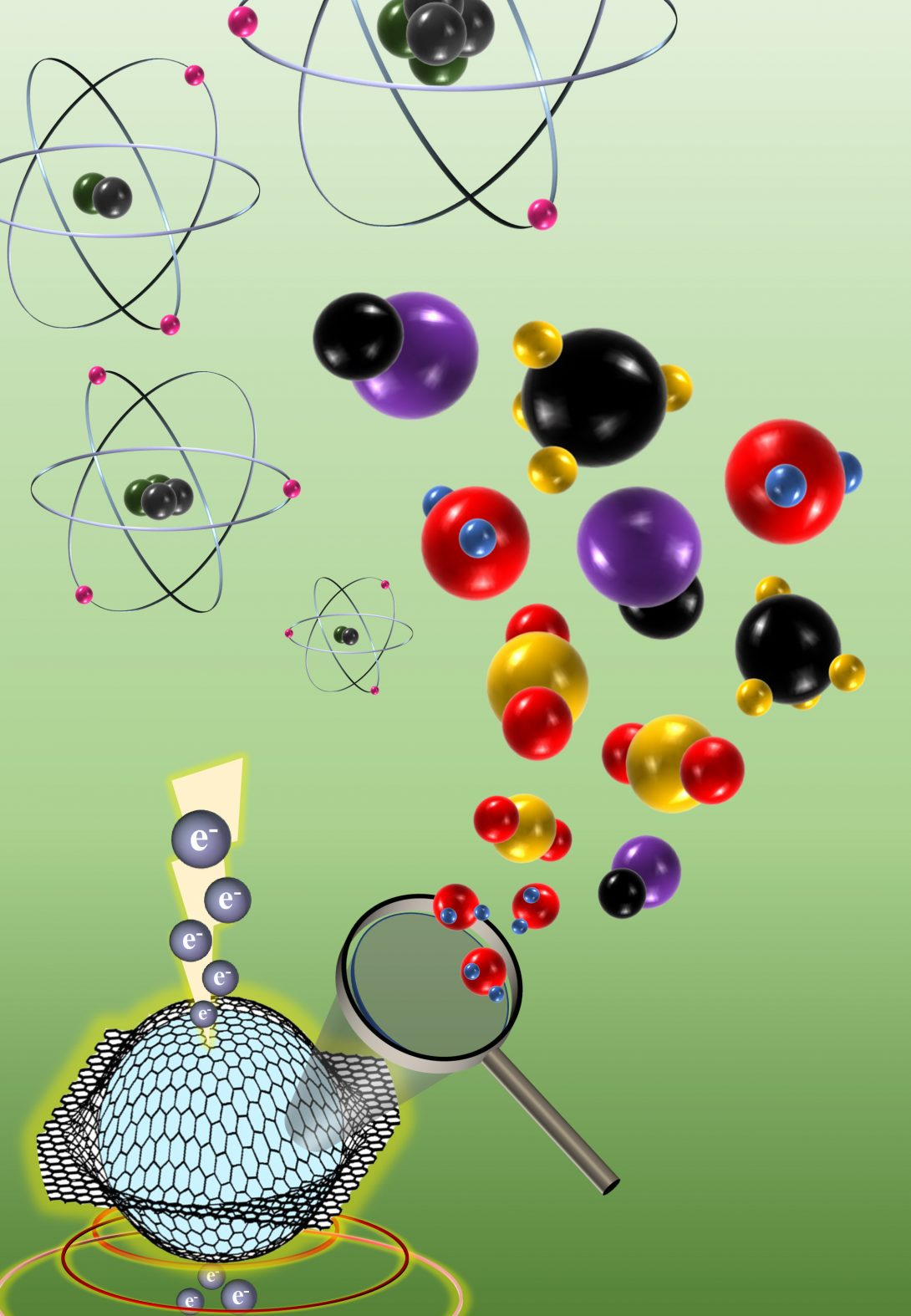
Imaging biological structures in liquid environment poses a significant challenge for conventional transmission electron microscopy (TEM) due to stringent requirements of ultra-high vacuum design and narrow pole piece gap for sample insertion in the microscope column. Although recent developments in silicon-based microfluidics have enabled the flow of liquids between thin silicon nitride membranes for in-situ TEM experiments, this technique suffers from low spatial and analytical resolutions for imaging and spectroscopy. The most recent liquid-cell TEM technique, graphene liquid-cell (GLC) microscopy, employs only layers of graphene to encapsulate liquid specimens. Recent efforts with GLC-TEM have demonstrated superior imaging resolution and spectroscopic analysis of beam-sensitive specimens.
Seyed Ghodsi, a bioengineering Ph.D. candidate at In Situ Nanomedicine Laboratory (ISNL), has reviewed the parameters that affect the quality of GLC imaging and analysis in his most recent publication in Small Methods journal titled “Advances in Graphene-Based Liquid Cell Electron Microscopy: Working Principles, Opportunities, and Challenges.” Seyed has been working on the state-of-the-art in situ electron microscopy techniques that enable future scientists to observe biological samples with superior resolutions, something that is the specialty of Tolou Shokuhfar (BioE) and Reza Shahbazian-Yassar (MIE) groups.
In this review article, several important factors that affect the in-situ imaging of biological specimen, including the variations in GLC geometries and capillary pressure are discussed. The interaction between the electron beam and the sample, along with the possibility for artifacts or the formation of radical ions in the GLC are also highlighted. The scientific discoveries enabled by GLC-TEM in the areas of biomineralization, corrosion, as well as high-resolution imaging of organelles and proteins are briefly discussed in this review. Finally, the possible future research directions of GLC-TEM and the associated challenges have been addressed.
The application of GLC-TEM in areas such as materials science, life sciences, electrochemistry, and environmental sciences. 3D Reconstruction of Nanoparticles: Platinum nanoparticle assembly and transient morphology in solution (© 2015 Science Publications).[1] 3D Motion of Proteins: TEM observation of gold-conjugated DNA molecules in water (© 2013 American Chemical Society).[2] Protein Elemental Mapping: EELS maps of oxygen, iron and nitrogen of ferritin suspension in GLC (© 2014 Wiley Publications).[3] Solid-Electrolyte Interphase Formation: SEI layer formation in Li-ion batteries (© 2016 Elsevier Publications).[4] Water State Transition: Observation of water crystal formation upon entrapment in capillaries (© 2015 Nature Publications).[5] Nanoparticle Nucleation: Nucleation, precipitation and grain boundary formation of Thenardite in aqueous phase (© 2015 American Chemical Society).[6]
references Heading link

References
[1] J. Park, H. Elmlund, P. Ercius, J. M. Yuk, D. T. Limmer, Q. Chen, K. Kim, S. H. Han, D. A. Weitz, A. Zettl, A. P. Alivisatos, Science (80-. ). 2015, 349, 290.
[2] Q. Chen, J. M. Smith, J. Park, K. Kim, D. Ho, H. I. Rasool, A. Zettl, A. P. Alivisatos, Nano Lett. 2013, 13, 4556.
[3] C. Wang, Q. Qiao, T. Shokuhfar, R. F. Klie, Adv. Mater. 2014, 26, 3410.
[4] J. Y. Cheong, J. H. Chang, H. K. Seo, J. M. Yuk, J. W. Shin, J. Y. Lee, I. D. Kim, Nano Energy 2016, 25, 154.
[5] G. Algara-Siller, O. Lehtinen, F. C. Wang, R. R. Nair, U. Kaiser, H. A. Wu, A. K. Geim, I. V. Grigorieva, Nature 2015, 519, 443.
[6] J. M. Yuk, Q. Zhou, J. Chang, P. Ercius, A. P. Alivisatos, A. Zettl, ACS Nano 2015, 10, 88.