BME undergrad updates testing device to improve biocompatibility of medical implants
block 1 Heading link
Biomaterials must undergo strenuous material testing to determine the safety or biocompatibility of the device in environments such as the human body. An undergraduate student in the Richard and Loan Hill Department of Biomedical Engineering has created a cost-effective device that could help researchers improve the safety of these vital treatments.
The biocompatibility, which is the ability of a medical device to perform with an appropriate host response in a specific application, and osseointegration as well as the corrosion and tribocorrosion properties of the material must be fully evaluated when developing novel biomaterials.
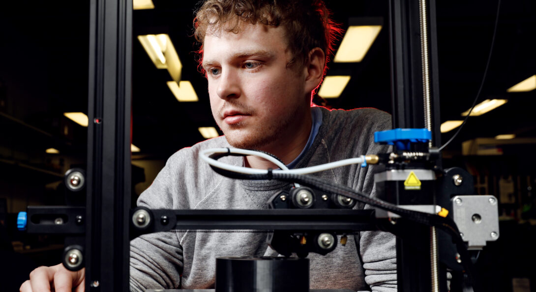
Looking to improve the material properties of implants to make them more durable, reduce failure rates, and improve function, undergraduate student Kyle Kinnerk developed an updated corrosion cell device.
Corrosion cells test the reaction of metal specimens in a harsh environment, such as an implant in the human body. The 3D-printed cell is made of polysulfone, a material that can withstand highly heated chemical reactions, which allows for cell culture and toxicity studies.
“The danger of an implant corroding in the human body is that, in cases of failure, small metal particles will fall off the implant and will be released into the surrounding tissue that can cause bone resorption and cancerous tumors,” Kinnerk said. “If these metal particles get into the bloodstream, they can spread throughout the body instead of remaining localized to the area where the implant is and increase the risk of metal toxicity to remote organs.”
Biomedical implants are a widely accepted clinical solution to severe arthritis and other health issues in orthopedics and dentistry. The failure rate of medical implants such as hip replacement implants over a period of 10 years is about 4% and yet, 1 in 10 Americans have a metal-based implant in their body.
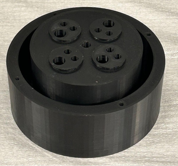
After joining Professor Mathew Mathew’s lab, Kinnerk designed the device to allow up to four separate biomaterial accelerated corrosion experiments to occur simultaneously. Using a corrosion cell, researchers can simulate the degradation in the implants over time in the body in short-duration experiments.
CAD modeling and 3D printing helped Kinnerk enhance the device’s design lessening the material usage, weight of the device, and the time required for manufacturing. As the corrosion science market is somewhat niche, corrosion cells typically cost almost $3,000 per unit, although Kinnerk’s new design costs only $20 to $50 per unit.
“One of the biggest advantages of the new manufacturing method is in the ability to prototype further and push this technology in new directions, whereas previous manufacturing was too costly and too limited to allow us to really prototype the device,” Mathew said.
Previously, the cost prohibited the corrosion cell from becoming a widely adopted technology. However, the implementation of 3D printing to the design of the corrosion cell will allow for further research to be conducted in the field of biomaterials to improve the material properties of dental implants, orthopedic implants, and other medical implants to improve patient outcomes.
Material and Civil Engineering PhD student Yani Sun contributed to this project.