UIC professor advances safer, more sensitive brain imaging for newborns
block
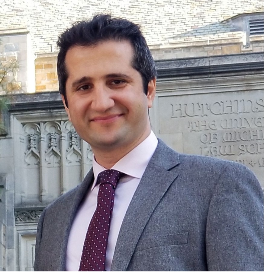
Each year, thousands of premature babies face a silent and dangerous threat: brain hemorrhages that can lead to long-term neurodevelopmental issues. Detecting these tiny bleeds early can make all the difference — but current imaging techniques are limited.
At the University of Illinois Chicago, Professor Kamran Avanaki from the Richard and Loan Hill Department of Biomedical Engineering is pushing the boundaries of what’s possible in neonatal brain imaging.
A window into the brain
In newborns, particularly those born prematurely, doctors often use ultrasound through the fontanelle—the soft spot on a baby’s head—to look for internal brain bleeding. It’s the safest and most practical option for fragile infants, as alternatives like MRI require sedation and moving the baby out of the neonatal unit.
But there’s a problem with this approach.
“Ultrasound is helpful, but it often can’t detect small blood clots,” Avanaki explains. “You need a certain amount of bleeding before it shows up. That delay in detection could cost precious time.”
According to the National Institutes of Health, 20–25% of babies born before 30 weeks or weighing under 1500 grams experience intracranial hemorrhages, and early, accurate detection is critical.
Lighting the way with photoacoustics
To address this gap, Avanaki and his team turned to photoacoustic imaging, a cutting-edge technique that uses laser pulses instead of sound waves. When the laser enters the tissue, molecules like hemoglobin absorb the light and generate tiny ultrasound signals. These signals help detect even small traces of blood, revealing early signs of hemorrhage that traditional ultrasound might miss.
Building on this success, Avanaki asked a bold question: Can we go even deeper?
Microwave energy, clearer insight
That curiosity led to the development of thermoacoustic imaging. Instead of light, it sends low-energy microwave pulses into the brain. These waves travel deeper than light and produce acoustic signals when absorbed — similar to photoacoustic imaging — and are detected using familiar ultrasound receivers.
“In both methods, we’re receiving ultrasound signals,” Avanaki says. “But in photoacoustics, we send light. In thermoacoustics, we send microwaves. The beauty is, we can use similar handheld probes already familiar to physicians, and the entire process is bedside and noninvasive.”
From bench to bedside: testing the tools
The team validated the technology in a step-by-step process. First, they created brain-like tissue models with artificial hemorrhages. Then, they tested the devices on sheep heads from butcher shops. Finally, in collaboration with UIC’s Department of Neurosurgery, they created artificial fontanelles in live sheep models, mimicking human newborn conditions.
The result? Exceptional sensitivity in detecting even small hemorrhages, with the new technique outperforming traditional ultrasound.
“This has been one of the most rewarding projects I’ve worked on,” Avanaki said. “Not only because of the potential clinical impact, but also because it pushed us to keep innovating at every step.”
Collaboration at its core
Avanaki credits much of the project’s success to a deeply collaborative team, including Dr. Fady Charbel, Richard L. and Gertrude W. Fruin Professor and Head of Neurosurgery at UIC, and Dr. De-Ann Pillers, Dharmapuri Vidyasagar Professor of Neonatology, and Program Director of UIC’s Fellowship Training Program in Neonatal-Perinatal Medicine
Their combined expertise, from biomedical engineering to neurosurgery and neonatology, helped bring a visionary idea to life.
Professor Avanaki’s recent publications on this work have appeared in leading journals including, Photoacoustics, Scientific Reports, and IEEE Transactions on Medical Imaging.
As brain imaging for newborns continues to evolve, Avanaki’s work brings us closer to a future where fragile infants get faster, more accurate diagnoses and better outcomes from the very beginning of life.