Improving prostate cancer detection through MRE
Text block one Heading link

A non-invasive imaging test that measures the stiffness of tissue may help to improve the diagnosis of prostate cancer, UIC research shows. This is crucial for patients, as early and accurate diagnosis of the cancer leads to improved treatment results.
Richard and Loan Hill Department of Bioengineering Associate Professor Dieter Klatt and colleagues recently published their research into how magnetic resonance elastography, or MRE, provides critical information for prostate cancer diagnosis.
Klatt said the current procedure for detecting prostate cancer involves a health care provider manually checking the mechanical properties of the prostate along with some use of magnetic resonance imaging, or MRI.
“We wanted to show that MRE can provide valuable information for in vivo prostate cancer diagnosis,” Klatt said.
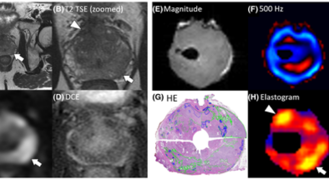
To accomplish this, the group performed MRE on prostate tissue and tested the MRE results against findings of pathology.
“Excitingly, we found high specificity of MRE for prostate cancer diagnosis, which has been a limiting factor of other diagnostic techniques,” he said. “In addition to increasing specificity, MRE provides quantitative measures which may reduce interreader variability of radiologists.”
MRE is an imaging technique that has been used since the mid-1990s to diagnose liver fibrosis. It measures the stiffness of tissue by sending mechanical waves through it and creating an elastogram, or stiffness map. In the case of prostate cancer, MRE can be used to guide surgeons in their efforts to remove cancerous tissue, which may help to reduce complications such as urinary and sexual dysfunction.
For this paper, Klatt and colleagues used 14 fresh prostate specimens from men with clinically significant prostate cancer who had undergone surgery but not radiation therapy. They used MRE to map the cancerous and healthy tissue in the specimens. The paper, titled “Prostate cancer assessment using MR elastography of fresh prostatectomy specimens at 9.4 T,” was published in Magnetic Resonance in Medicine.
The group is investigating whether MRE can be used to detect other illnesses, such as Alzheimer’s disease.
“In a mouse study of Alzheimer’s, we found that in a neurodegenerative brain, the tissue stiffness decreases,” Klatt said. “Interestingly, this is opposite to prostate cancer and fibrotic diseases, where the tissue becomes stiffer with disease.”
Additional authors on the Magnetic Resonance in Medicine paper include Rolf Reiter, Shreyan Majumdar, Steven Kearney, Andre Kajdacsy‐Balla, Virgilia Macias, Simone Crivellaro, Brandon Caldwell, Michael Abern, and bioengineering department head Thomas Royston.